Results
This page presents current findings from our test scans and explores what EDI may be capable of identifying. To date, 40 volunteer subjects (31 female, 9 male) have participated in EDI scans. Most had no known medical conditions, while a few did. Scans covered various body regions—some recorded once, others multiple times. This page highlights a selection from over 200 collected scans.
Important Disclaimer: The results shown on this page do not imply that EDI has diagnostic capability. This will be subject to rigorous testing and evaluation through formal clinical trials. Any views, comments, or interpretations presented here should be considered exploratory and not as medical fact. If you have concerns about your health, please consult a qualified medical practitioner.
Women’s Health
Women’s health has been a key focus area, especially due to the prevalence of breast cancer. Early findings suggest EDI may have potential applications in breast health—including screening, cancer detection, and supporting existing imaging techniques. It may even offer advantages for examining younger individuals with denser breast tissue.
EDI is safe and repeatable without health risks. In contrast, mammography is typically limited to women over approximately 45, due to reduced efficacy in dense breast tissue. It’s also contraindicated during pregnancy and cannot be repeated frequently due to cumulative radiation exposure.
EDI could complement and extend current screening programs. It’s suitable for all ages, including young women and those who are pregnant, and can be repeated without risk. It is painless for most subjects and entirely non-harmful. Preliminary studies also suggest EDI may reflect activity related to other aspects of women’s health—such as the ovaries and uterus. With further investigation, it may support research into reproductive cycles and gynaecological conditions.
Typical Frontal Female Scan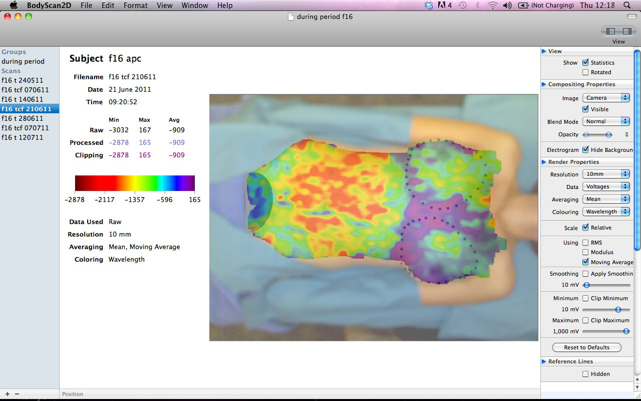 No history of breast conditions. One child, breastfed; no use of personal contraceptives. |
Post Lumpectomy of Breast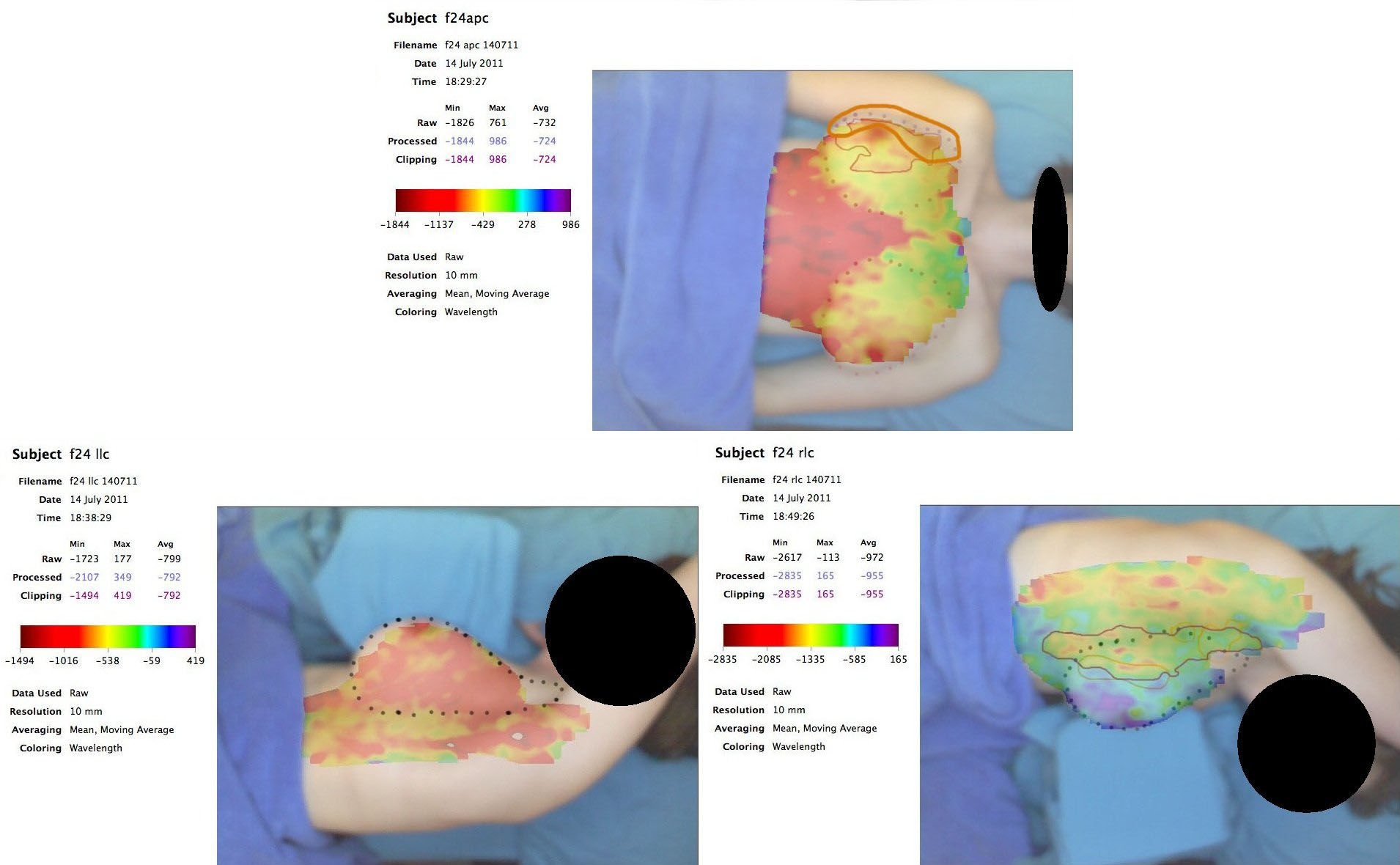 Lateral views show notable voltage differences between left and right breasts. Regions of surgical intervention are clearly delineated on the right. |
Breast Reconstruction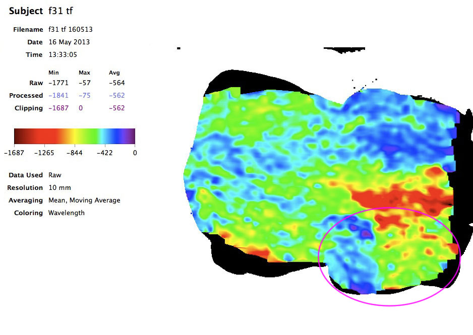 Left breast reconstruction using the TRAM procedure several years prior. Voltage patterns reveal similarities to abdominal tissue in the upper region, and alignment with the right breast in the lower region. |
PCOS – Ovarian Cyst Left Side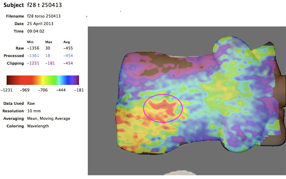 The circled region corresponds to the subject’s reported pain. The scan shows a distinct low-voltage zone in the same area. |
Cyclic Correlation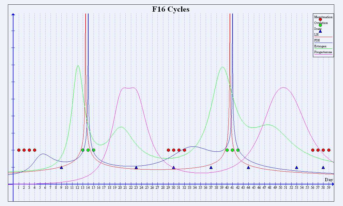 This chart from a female subject not using hormonal contraceptives suggests a potential cyclic correlation between simulated progesterone levels and minimum voltage readings at each scan. |
Cysts In Right Breast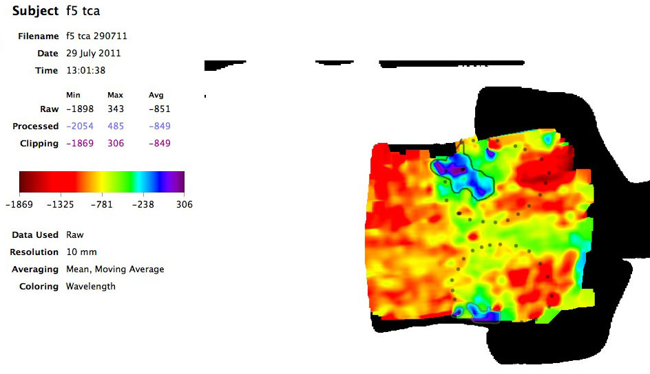 Cysts present in the right breast prior to fluid drainage. Higher voltages are observed over cysts compared to surrounding breast tissue. |
Soft Tissue
Our earliest tests focused on the back, where initial scans produced patterns that appeared to outline the lungs. Similar responses were observed in other subjects, prompting further exploration of additional body regions.
Over the course of EDI research, we've seen results that suggest potential value in examining soft tissue and joint conditions. Beyond medical diagnostics, EDI could have applications in sports and the performing arts—domains where soft tissue and joint injuries are common and rapid recovery is vital.
The back and legs are particularly injury-prone. For professionals whose careers depend on physical performance, identifying when to rest or locating problematic areas for targeted recovery is critical—something EDI may be able to support.
It may also help reveal injury extent and offer a more accurate method of tracking recovery and healing progress.
Typical Back Scan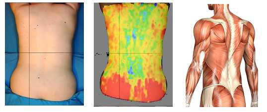 Electrical scan paired with anatomical reference. Distinct electrogram features show alignment with fascia and muscle distribution in the lower back. |
Possible Lung Detection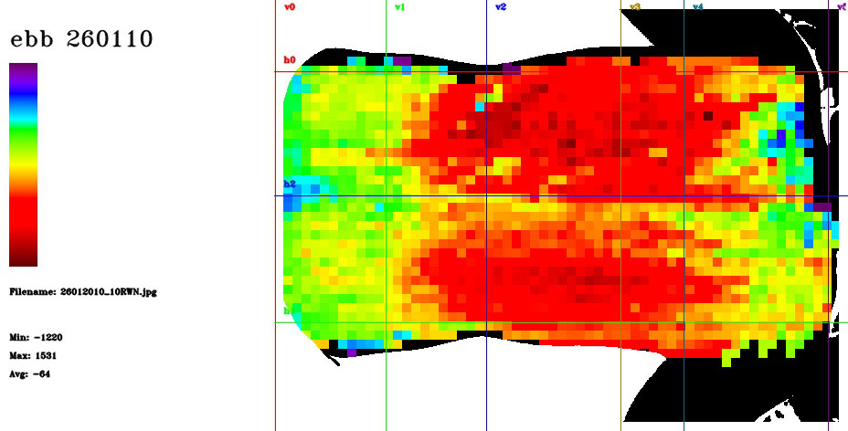 Scan from a healthy subject displaying two prominent low-voltage (red) regions. These may correspond to lungs or adjacent muscle/ligament structures. |
Shoulder Injury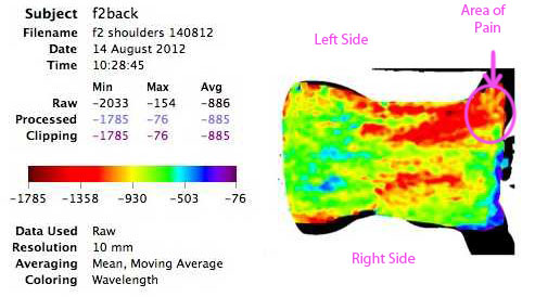 Back scan showing reduced voltage over the left shoulder, consistent with reported injury when compared to the right side. |
Spine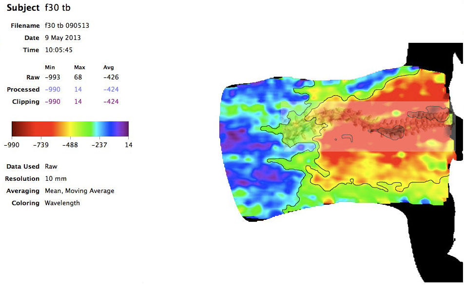 Scan showing scoliosis with overlay of spinal curvature. Multiple voltage zones highlight asymmetry and possible areas of muscle compensation. |
Scarring and Muscle Damage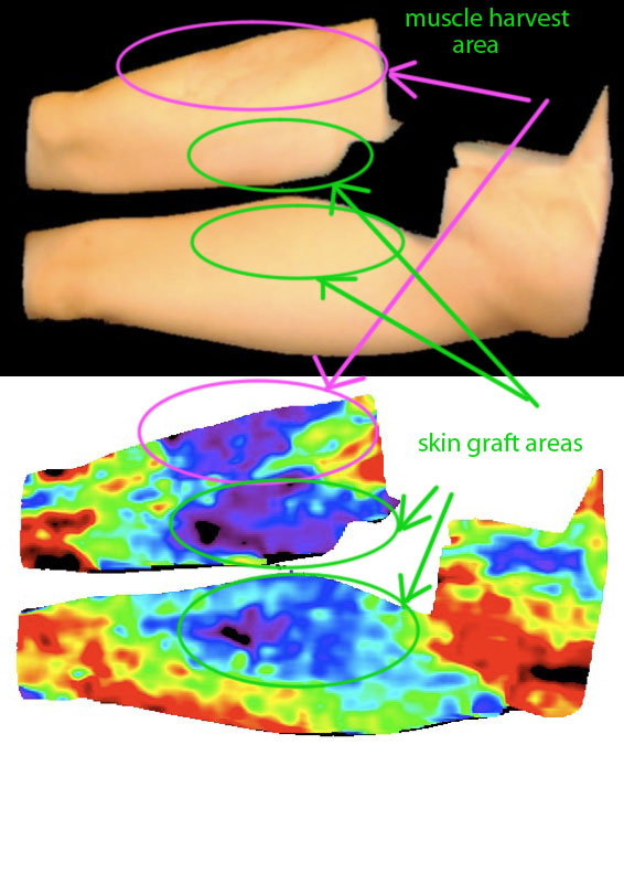 Post-operative scan following muscle harvesting and infection. Higher voltages are noted over damaged tissue compared to the unaffected leg. Skin graft sites are also visible. |
Stab Wound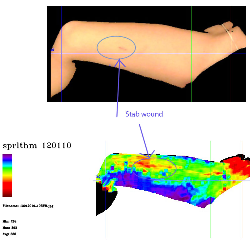 Scan taken two weeks post-injury. Lower voltages around wound site may reflect tissue trauma and ongoing healing processes. |
Other Results
Throughout our research, EDI scans have produced responses that may correspond to a variety of internal organs and structures. While still under investigation, these observations point toward potential applications across diverse areas of medicine. Alongside more consistent results, we’ve also encountered several isolated or unexpected scan effects that could merit further study.
Large Intestine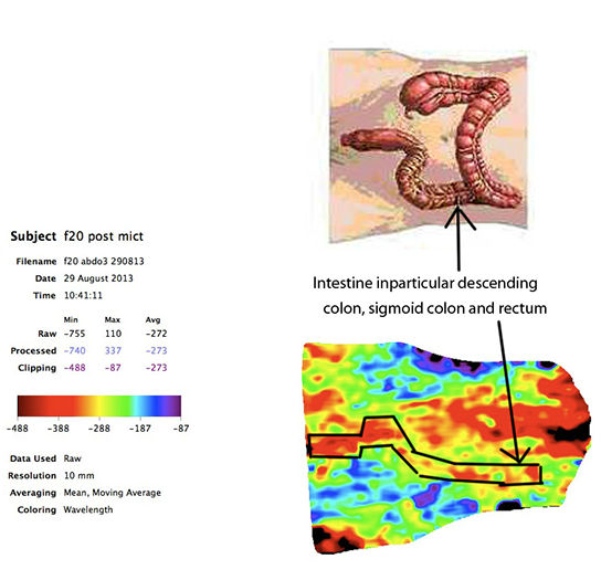 Scan patterns appear consistent with activity in the descending colon, sigmoid colon, and rectum. Subject bowel movement confirmed findings post-scan. |
Kidney Transplant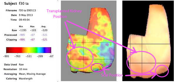 Electrical mapping indicates boundary responses around an implanted kidney. Circled areas show kidney placement and associated skin scarring. |
Kidney Stones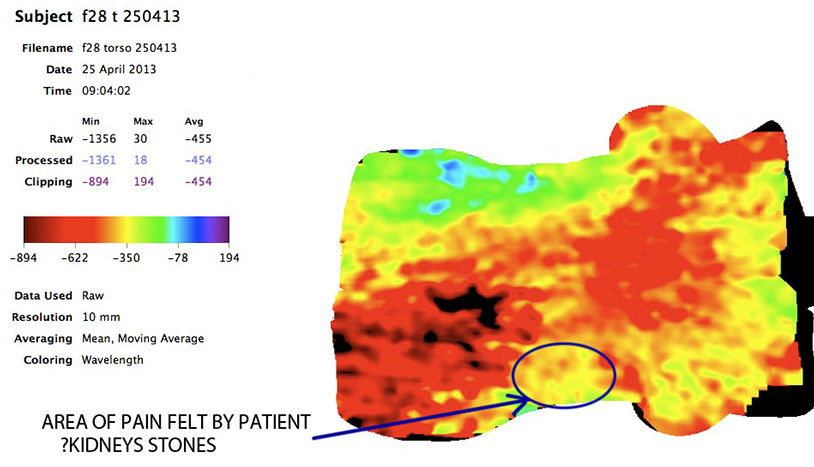 Scan from a subject with confirmed left kidney stones. Circled region aligns with reported pain and shows distinct voltage variation. |
Incontinence Issues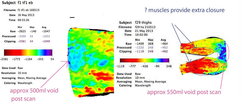 Left image shows full bladder presenting as a high-voltage zone. Right scan from subject with urge incontinence reveals reduced quadriceps voltage, possibly indicating muscular response. |
Wilson's Disease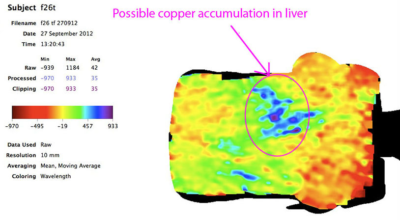 Subject undergoing Parkinsonism treatment displayed an unusual liver scan pattern, potentially suggestive of Wilson’s Disease—though unconfirmed and not previously screened. |